Patient Case
Patient History:
- 72 year old male
- Status post endovascular aortic repair (EVAR), followed by a thoracic endovascular repair (TEVAR) a year later
- Empyema found in the left lung and pleural cavity
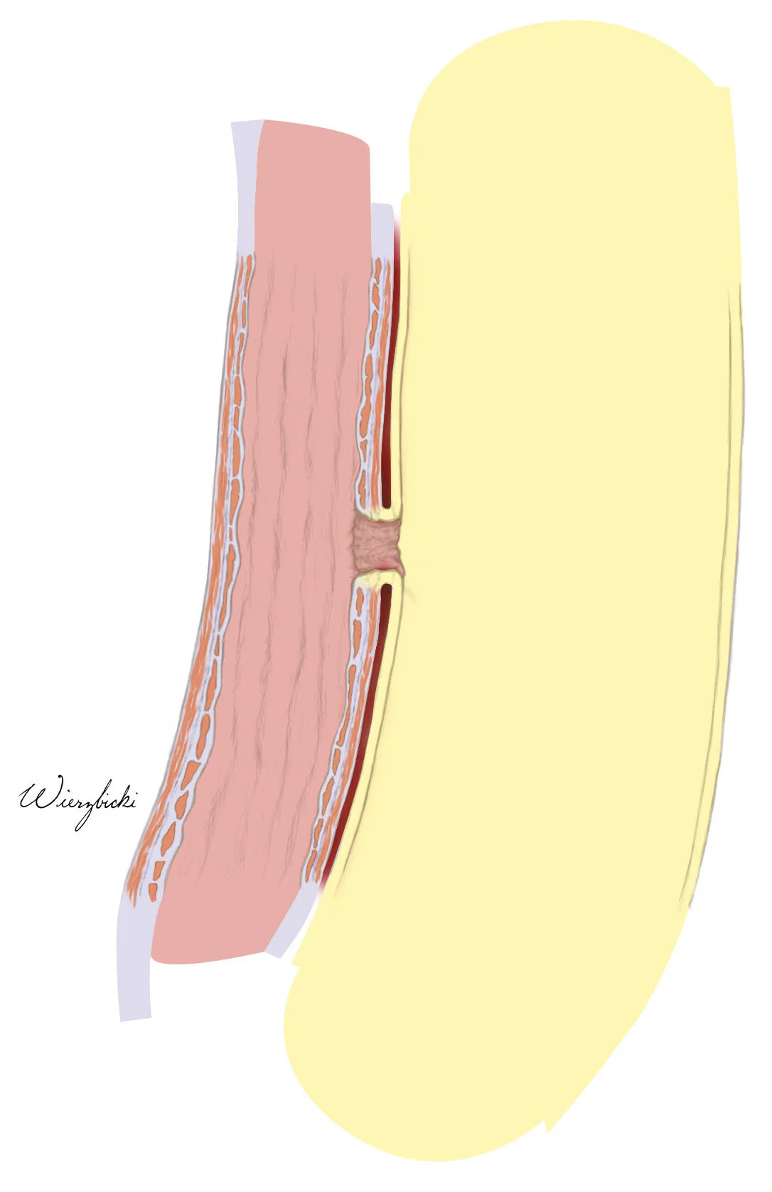
- Aortoesophageal fistula discovered due to an infected TEVAR
Sketches
Because Baylor College was not supplied with a full body scan of the patient’s aorta, I resorted to combining two different sets of CT scans – one of the patient’s thorax and another of their abdomen – to reconstruct the patient’s aorta via CT scan segmentation using Photoshop and OsiriX. There was a total of about 150 scans to go through for each set. The structures that were tracked include the esophagus, aorta, the arteries coming off the aortic arch, the celiac trunk, SMA, and renal arteries.
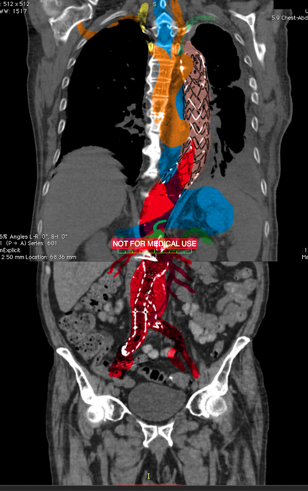

Different colors were used to identify and differentiate the structures that were being tracked through Photoshop.

A line sketch was created using the outlines of the blocked-off silhouette. Because the aortic valve was blocking the fistula from view, it was necessary to shift the valve and part of the ascending aorta slightly to the left so that it would be visible.
Rendering